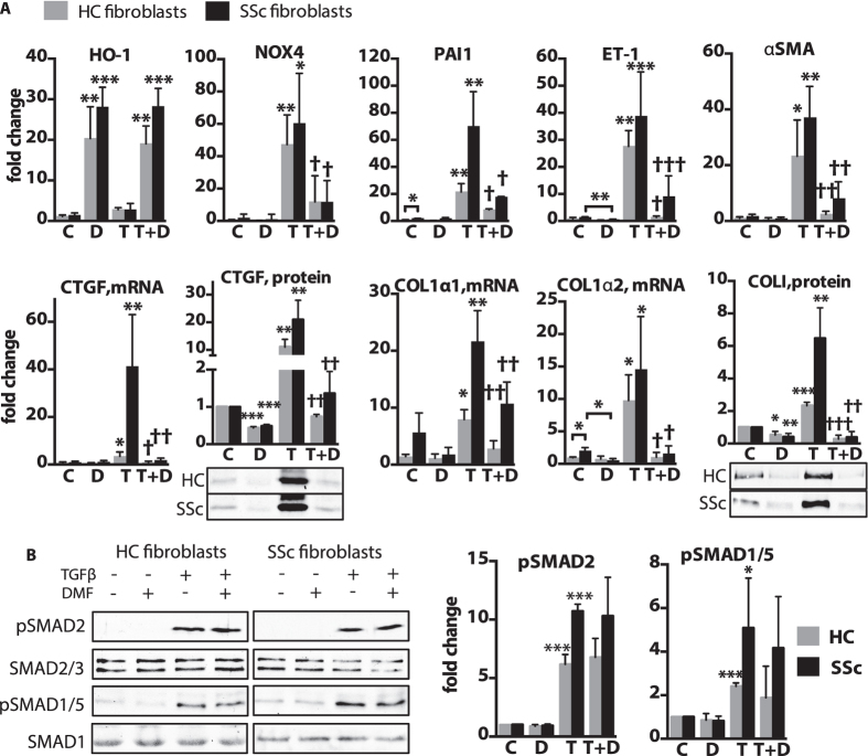
Figure 6. TGFβ –induced lung fibroblast activation is inhibited by DMF.
(A,B) Human primary lung fibroblasts from healthy controls (HC) and scleroderma patients (SSc) were treated for (A) 24 h with 2.5 ng/ml TGFβ(T) and 90 μM DMF(D), control cells (C) were treated with vehicle (DMSO), or (B) pre-treated for 1 h with DMF(D) and then exposed to TGFβ(T) for 30 min. (A) Relative gene expression was measured with qPCR. CTGF and COLI proteins were measured by densitometry of immunoblots from the whole cell lysates. (B) Representative immunoblots and densitometry measurements of pSMAD2 and pSMAD1/5 levels. n = 3 cell lines per group. Data shown as mean ± SD. *P versus control (C); †P versus TGFβ(T). *P < 0.05, **P < 0.01, ***P < 0.001. The uncropped images of selected immunoblots are shown in Supplementary Fig.14.

