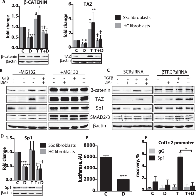
Figure 7. DMF promotes βTRCP-mediated degradation of Sp1, β-catenin and TAZ.
(A,D) Lung fibroblasts from patients were treated for 24 h with TGFβ(T) and DMF(D) or vehicle (C). Representative immunoblots and densitometry measurements of β-catenin and TAZ. n = 3 cell lines per group. (B) Lung fibroblasts (IMR90) were pre-treated with proteasome inhibitor MG132 for 1 h and then exposed to TGFβ with DMF or vehicle for 24 h. (C) IMR90 were silenced with scrambled siRNA (SCRsiRNA) or βTRCP siRNA and then treated with TGFβ with or without DMF treatment for 24 h. Representative immunoblots are shown. (D) Representative immunoblots and densitometry measurements of Sp1. (E) Lung fibroblasts were transfected with COL1α2 promoter reporter and then treated for 24 h with DMF or vehicle. n = 3 independent experiments. (F) Chromatin immunoprecipitation using Sp1 antibody was performed after 24 h treatment with TGFβ and DMF or vehicle. The amount of recovered DNA was measured with qPCR. n = 3 independent experiments. Data shown as mean ± SD. *P versus control (C); †P versus TGFβ(T). *P < 0.05, **P < 0.01, ***P < 0.001. The uncropped images of selected immunoblots are shown in Supplementary Fig. 14.

