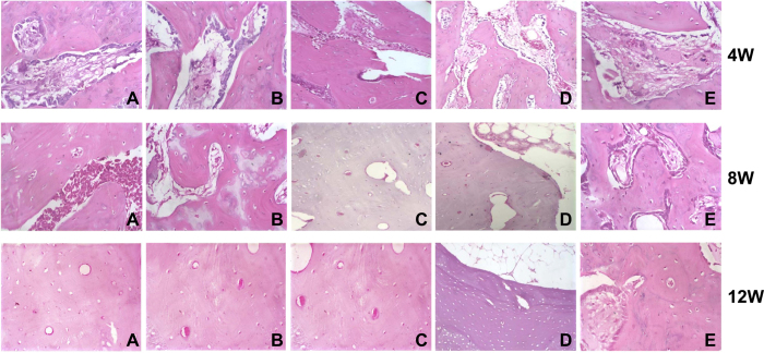
Figure 6. Histological observation of osteogenesis among the five groups at the 4th, 8th and 12th weeks.
Note: (A) in vitro prefabrication with EPCs-RFP + in vivo prefabrication with arteriovenous vascular bundle + secondary osteoblast (OB)-GFP implantation; (B) in vitro prefabrication with EPCs-RFP + secondary OBs-GFP implantation; (C) in vivo prefabrication with arteriovenous vascular bundle + secondary OBs-GFP implantation; (D) implantation of EPCs-RFP + implantation of with arteriovenous vascular bundle + simultaneous OBs-GFP implantation; (E) only DBM grafts (blank control); ECs, endothelial cells; OB, osteoblast; RFP, red fluorescence protein; GFP, green fluorescence protein.

