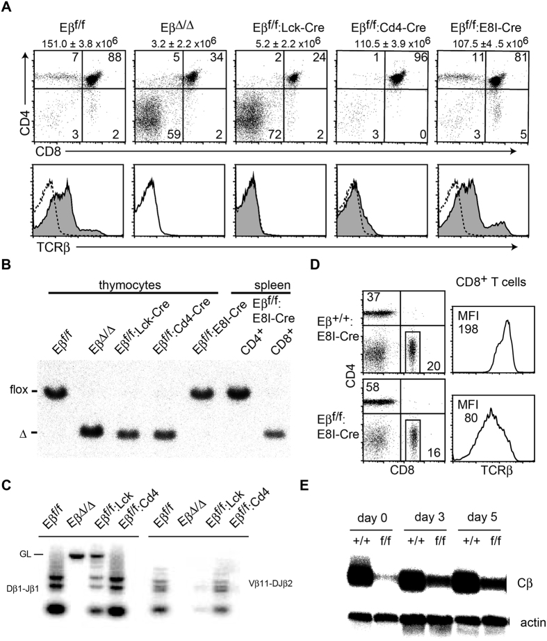
Figure 3. Effect of conditional deletion of Eβ at distinct developmental stages on TCRβ expression.
(A) Expression levels of CD4, CD8 and TCRβ on total thymocytes from Eβ f/f, EβΔ/Δ, Eβ f/f:Lck-Cre, Eβ f/f:Cd4-Cre and Eβ f/f:E8I-Cre mice are shown. TCRβ expression in EβΔ/Δ mice is shown as a dotted line in the histogram as a control. (B) DNA from total thymocytes and sorted CD4+ and CD8+ splenocytes from mice with the indicated genotype was analyzed for the efficiency of Cre-mediated deletion of Eβ by Southern blot. The bar indicates the position corresponding to the Eβ f and EβΔ allele. (C) DNA-PCR analyses for analyzing Dβ1-Jβ1 and Vβ11-DJβ2 recombination in sorted CD25+CD44− DN3 thymocytes from indicated mice. The bar indicates the position corresponding to the germline configuration. (D) Expression levels of TCRβ on CD8+ lymph node cells from Eβ +/+:E8I-Cre and Eβ f/f:E8I-Cre mice are shown as histograms with mean fluorescent intensity (MFI) at the left upper corner. (E) Northern blot showing TCRβ transcripts detected by the Cβ probe. CD8+ splenocytes from Eβ +/+:E8I-Cre and Eβ f/f:E8I-Cre mice were activated by TCR stimulation. Total RNA was prepared before and three or five days after stimulation. Five micrograms of total RNA was loaded in each lane. The actin mRNA was used as the loading control.

