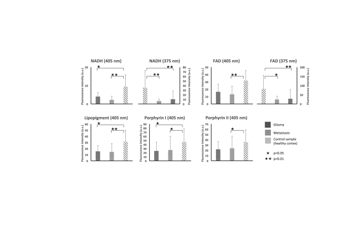
Figure 1. Results of spectroscopic endogenous fluorescence measurements for intra-axial tumor part.
Fluorescence intensity of NADH and FAD when exciting with 405 and 375 nm for Glioma, metastasis and control samples. Results for Lipopigment, porphyrin I and II are presented for 405 nm excitation wavelength. ★★Under a bar denote statistically significant difference (p < 0.01).

