
Figure 5. Lack of significant changes in the expression of GAD65 in the VB of inflammatory pain rats.
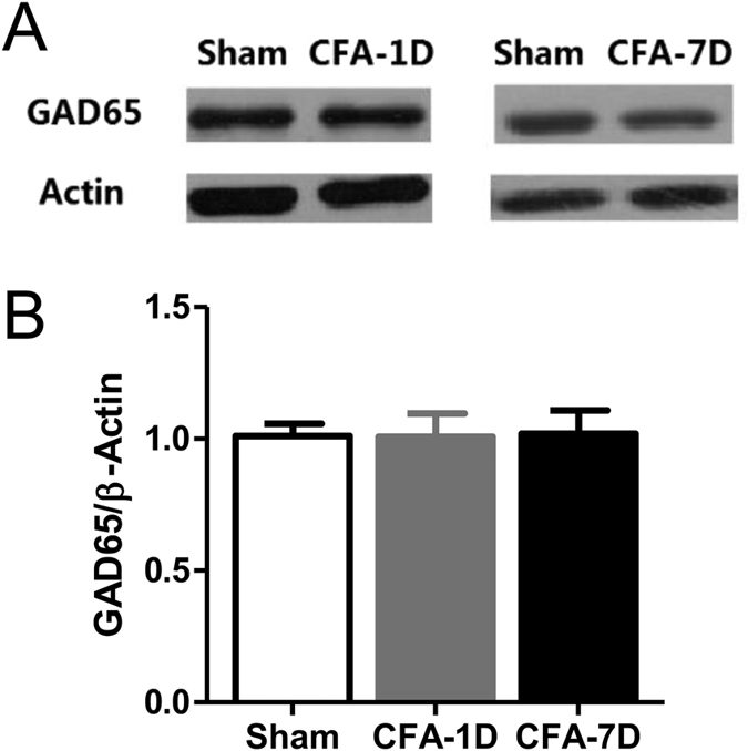
(A) Representative Western blotting bands of GAD65 (~65 KD). β-actin was used as the reference. (B) Statistics of relative quantification of GAD65 in the control, CFA-1D (day 1 after CFA injection) and CFA-7D (day 7 after CFA injection) groups. Error bars indicated SEMs, unpaired t test.