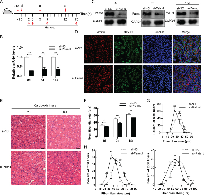
Figure 5. Knockdown of Palmd in vivo impairs muscle regeneration.
(A) Schematic representation of CTX injury followed by si-NC or si-Palmd treatment in mouse TA muscle. (B) The mRNA level changes of Palmd in TA muscle at 2d, 7d and 15d, after CTX injury (−1d) and siRNA injection (0d, 5d and 12d). (C) The protein levels of Palmd in TA muscle of si-NC or si-Palmd group on 3d 7d and 15d. (D) Immunofluorescence staining for Laminin (red), eMyHC (green) and nucleus (blue) on si-NC or si-Palmd group TA muscle section on 3d. Scale bar = 75 μm. (E) H&E staining on si-NC or si-Palmd treated TA muscle section at 7d and 15d. Scale bar = 50 μm. (F) TA muscle average myofiber diameters (μm) of si-NC or si-Palmd group at 3d, 7d and 15d. n = 3 per group, more than 300 myofibers per mouse. (G~I) Percent distributions by diameter (μm) of myofibres at 3d (G), 7d (H) and 15d (I) analyzed in (F). Data are presented as mean ± s.e.m., n = 3 per group. **p < 0.01, ***p < 0.001.

