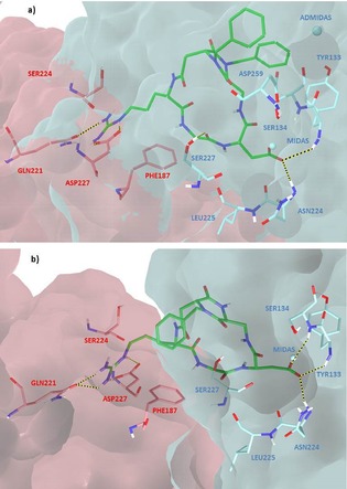
Figure 6.

Docking best poses of a) compound 7 (green) and b) compound 6 (green) in the crystal structure of the extracellular domain of α5β1 integrin (α5 subunit pink, β1 subunit cyan, model from 3VI4.pdb). Only selected integrin residues involved in interactions with the ligand are shown. Non polar hydrogens are hidden for clarity, whereas intermolecular hydrogen bonds are shown as dashed lines.
