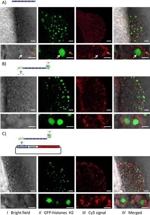
Figure 4.

Localization of the DONs in the developing zebrafish embryos. One‐cell stage embryos from a zebrafish line stably expressing GFP‐fused histone H2A were used,33 which allows the direct localization of the cell nuclei (green channel). The Cy5‐labeled constructs (red channel) were microinjected into the yolk with DON‐1Cy5 alone (A), the protein‐decorated DON (DON‐1Cy5‐GV; B) or DON‐1Cy5‐GV along with the reporter plasmid (C). Then, at 6 h post‐fertilization, the embryos were embedded in agarose and analyzed by using confocal fluorescence microscopy. Three channels were recorded: the bright‐field image (I), the signal from the GFP‐histone H2 in green (II), and the Cy5 signal from the DONs in red (III). The high‐intensity signal in the red channel shows that the large DNA constructs were internalized at a high rate from the yolk. In addition, the high magnification (bottom images) clearly indicates that the internalized DONs are mostly cytosolic (orange arrow), with very little to no nuclear localization (white arrow). Note that the presence of neither the proteins nor the reporter plasmid influenced the internalization or the subcellular distribution of the DONs. Scale bars: 30 μm and 10 μm for low and high magnification, respectively.
