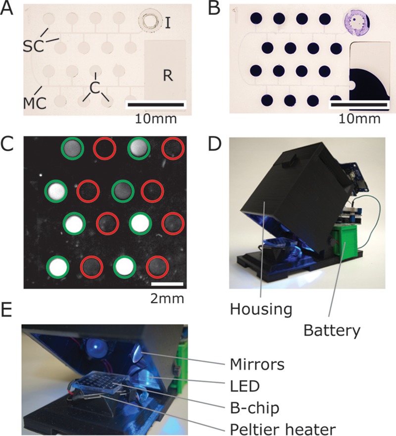
FIG 1.

Images depicting the B-chip and electronic reader. (A) An image of a B-chip containing 16 microchambers (C, each with a volume of 1 μl). (B) An image of a degassed B-chip that has been loaded with an aqueous solution of crystal violet (50 μl) at the inlet “I” after 20 min. Air fills the main and side channels (MC and SC, respectively), physically separates reactions, and prevents cross-contamination between the chambers. Excess sample is collected in the reservoir “R” (scale bar, 10 mm). (C) Representative image of fluorescence from on-chip RPAs for S. aureus, imaged with an ImageQuant; green circles indicate the presence of S. aureus-specific primers, and red circles indicate the absence of primers (scale bar, 2 mm). (D and E) Images of the B-chip reader highlighting the housing, optical components, and a Peltier plate to heat the sample.
