
FIG 2.
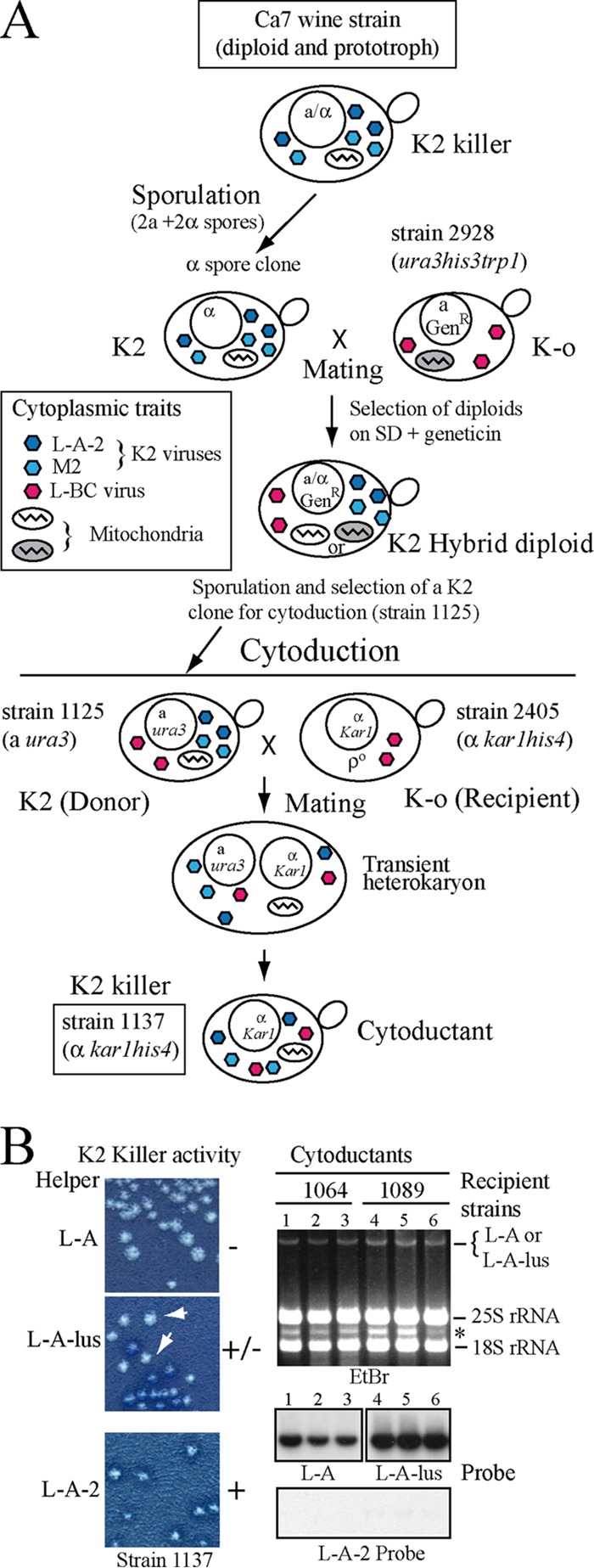
Construction of K2 strain 1137 and exclusion of K2 by L-A or L-A-lus. (A) Diagram of the experimental approach followed to introduce the K2 killer trait from wine strain Ca7 into laboratory strain 2405. The K2 strain Ca7 (diploid and prototroph) was induced to sporulate, and the spore clones were mated to laboratory strain 2928 (expressing Geneticin resistance [GenR] from a vector [23]). Hybrid diploids were selected on minimal medium (SD) with Geneticin. After sporulation, strain 1125 was selected (a ura3; K2). The cytoplasm of this K2 strain was then transferred by cytoduction into recipient strain 2405 (kar1 mutant defective in nuclear fusion and [rho0]). A transient heterokaryon was initially formed. After mitotic division, cytoductants carried the nucleus of the recipient strain and the mixed cytoplasms from the donor and recipient strains, and thus, they are K2 (strain 1137). Strain 1137 also carries L-BC virus (from strain 2928, since strain Ca7 is L-BC-o), and its mitochondria were inherited from the wine strain (information obtained from Fig. S5). The various cytoplasmic traits present in the cells are indicated as follows. K2 viruses L-A-2 and M2 are shown as dark-blue and light-blue hexagons, respectively; L-BC viruses are represented by red hexagons. Mitochondria from different parental strains are shown on a white or gray background. (B) K2 viruses are excluded by L-A or L-A-lus. L-A-2 and M2 viruses were transferred by cytoduction from strain 1125 into two strains isogenic with 2405 but containing L-A (strain 1064) or L-A-lus (strain 1089). (Left) (Top and center) K2 killer activity of isolated colonies from each strain, with the respective helper virus indicated. White arrows indicate colonies that have lost killer activity. (Bottom) K2 killer activity of strain 1137 carrying L-A-2 and M2, shown as a control. (Right) Total nucleic acids from three cytoductants from each recipient strain were separated in an agarose gel and were analyzed by Northern hybridization with probes specific for L-A (lanes 1 to 3) or L-A-lus (lanes 4 to 6). (Top) Ethidium bromide (EtBr)-stained gel. The band indicated by the asterisk corresponds to the 23S RNA narnavirus originally present in donor strain 1125. (Center) The two autoradiograms. (Bottom) The same samples (from lanes 1 to 6) annealed to the L-A-2-specific probe.