Fig. 2.

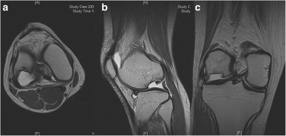
Preoperative magnetic resonance image. a Axial, b sagittal and c coronal images showed large osteochondral defect (approximately 2.7 cm × 2.2 cm sized and 1.5 cm deep) on lateral femoral condyle with osteochondral loose body

Preoperative magnetic resonance image. a Axial, b sagittal and c coronal images showed large osteochondral defect (approximately 2.7 cm × 2.2 cm sized and 1.5 cm deep) on lateral femoral condyle with osteochondral loose body