Fig. 6.

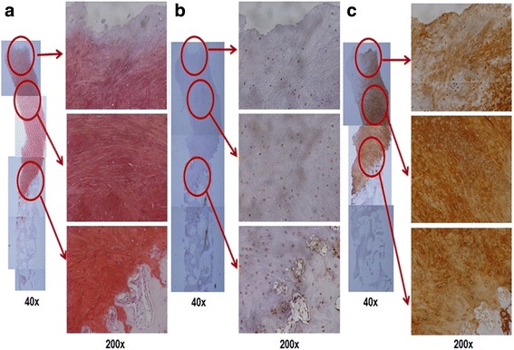
Histological findings. a Positive safranin – O staining was observed throughout the matrix. Immunostaining showed b weak staining for type I collagen but c diffuse strong positivity for type II collagen

Histological findings. a Positive safranin – O staining was observed throughout the matrix. Immunostaining showed b weak staining for type I collagen but c diffuse strong positivity for type II collagen