Figure 3.
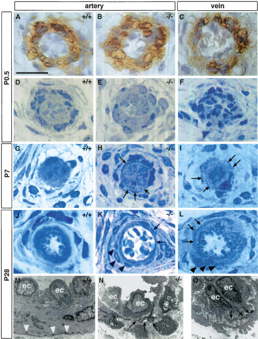
Impaired postnatal maturation of the lateral arteries from the tail in Notch3–/– mice. (A–C) α-SMA staining of P0.5 arteries from wild-type (A) and Notch3–/– mice (B) and vein from wild-type mice (C) showing that arteries and veins are surrounded by α-SMA-positive mural cells. (D–L) Toluidine blue staining of semithin sections at P0.5 (D–F), P7 (G–I), and P28 (J–L) from wild-type arteries (D,G,J), Notch3–/– arteries (E,H,K), and wild-type veins (F,I,L). At P0.5 wild-type and Notch3–/– arteries and wild-type vein appear similar. At later stages, vSMC in wild-type arteries harmoniously increase in length and thickness and become circumferentially oriented around the lumen, while vSMC in Notch3–/– arteries exhibit thin, irregular, and overlapping cytoplasmic processes (arrows), and form abnormal clusters of poorly oriented cells (black arrowheads). Note the similar aspect of vSMC from veins to those from Notch3–/– arteries at the same age. (M–O) Electron micrographs at P28 of wild-type artery (M), Notch3–/– artery (N), and wild-type vein (O), showing the irregular shape of vSMC with thin cytoplasmic expansions as well as the marked reduction of dense plaques (white arrowheads) in the mutant artery and the wild-type vein (ec, endothelial cells). Bars: A–L, 17 μm; M, 7.5 μm; N, 10 μm; O, 5 μm.

