Fig. 1.

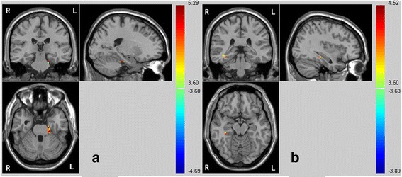
Compared with the control group, the SPD group exhibited decreased FC between the bilateral precuneus and opposite lateral (a: left; b: right) parahippocampus

Compared with the control group, the SPD group exhibited decreased FC between the bilateral precuneus and opposite lateral (a: left; b: right) parahippocampus