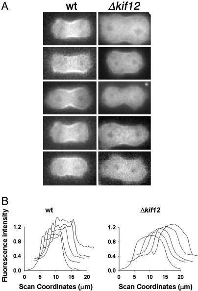
Fig. 4.
Observation of early furrowing in cells expressing GFP-myosin II. (A) Left shows five live wild-type (wt) cells undergoing cytokinesis. GFP-myosin II localization is observed prominently in the cleavage furrow. Right shows attempts of cytokinesis in live Δkif12 cells. The cells fail to localize GFP-myosin II to the cleavage furrow. (B) Line-scan analysis of fluorescence intensity of GFP-myosin II across the width of the cleavage furrow for each cell shown in A. The y axis shows the normalized fluorescence, and the x axis shows the scanning coordinate in micrometers.

