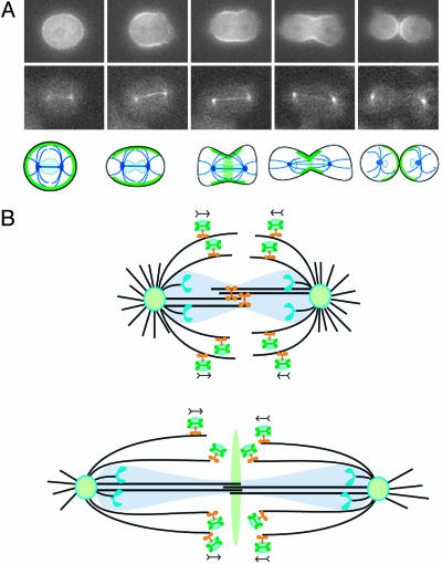
Fig. 5.
Progression of a live cell during cytokinesis and model for spindle-pole and vesicle movement during mitosis. (A) Top shows the localization and reorganization of myosin II during cytokinesis in a wild-type cell expressing GFP-myosin. Middle shows GFP-α-tubulin expressed in a wild-type cell under-going cytokinesis. Bottom is a schematic that shows the localization of myosin (green) and tubulin (dark blue) during cytokinesis in Dictyostelium wild-type cells. The position of the nuclei is indicated in light blue. (B) Upper shows Kif12 (orange) on microtubules (black) in the spindle region of a cell during anaphase. The nuclear membrane does not completely break down during mitosis of Dictyostelium and is shown in transparent light blue. The poles (light-green ovals) are separated, in part, by the action of Kif12 on the interdigitating central spindle microtubules. Kif12 also transports vesicles (light-blue ovals) carrying mechanoregulatory factors, perhaps including myosin II (green), on the spindle fibers. Chromosomes (blue) are being segregated bound to kinetochore microtubules. Lower shows a cell in late telophase. The vesicles drop off in the center of the cell and transfer the signals for cytokinesis.

