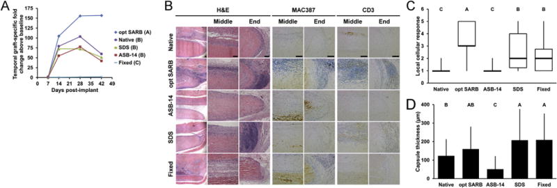
Fig. 2.

Systemic humoral, local innate and adaptive cellular, and local foreign body in vivo leporine responses to subpannicular BP implants. Temporal fold-increase in circulating graft-specific IgG titer post-implantation of BP implants (A). Representative images of BP implants at 42 d on H&E staining taken at low magnification (column 1), scaffold middle (column 2), and scaffold end (column 3). Representative images of MAC387 (macrophage, columns 4 and 5) and CD3 staining (T-cell, columns 6 and 7) staining taken at the middle and end of the scaffold at 42 d, respectively. Scale bar = 100 μm. Local cellular response as observed on H&E staining (C). Fibrous capsule thickness around BP implants at 42 d (D). Results plotted in (A) as mean (n = 6 per group and timepoint); (C) as mean ± standard deviation (n = 6 per group); and (D) as median (denoted by the thick line), inter-quartile range (25th–75th percentile), maximum (top whisker) and minimum (bottom whisker). Groups not connected by same letter are significantly different (p < 0.05).
