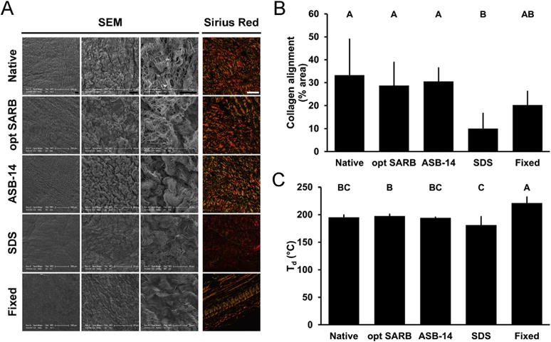
Fig. 3.

ECM architecture (morphology, collagen alignment, and thermal stability) of BP biomaterials. Representative images of BP biomaterials on scanning electron microscopy (serous side, columns 1–3) and picrosirius red staining under polarized light (column 4) (A). Scale bar = 100 μm unless otherwise stated. Collagen alignment of BP biomaterials observed on picrosirius red staining and expressed as percent area of field of view (B). Denaturation temperature (Td) of BP biomaterials (C). Results plotted as mean ± standard deviation (n = 6 per group). Groups not connected by same letter are significantly different (p < 0.05). (For interpretation of the references to color in this figure legend, the reader is referred to the web version of this article.)
