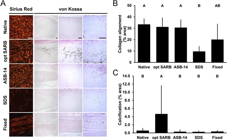
Fig. 4.

Collagen alignment and adaptive immune response-mediated in vivo calcification of leporine subpannicular BP implants. Representative images of 42 d BP implants on picrosirius red staining under polarized light (column 1) and von Kossa staining (Columns 2–4) (A). Scale bar = 100 μm. Collagen alignment of BP implants observed on picrosirius red staining and expressed as percent area of field of view (B). Adaptive immune response-mediated calcification of BP implants observed on von Kossa staining and expressed as percent area of field of view at sites of calcification (C). Results plotted as mean ± standard deviation (n = 6 per group). Groups not connected by same letter are significantly different (p < 0.05). (For interpretation of the references to color in this figure legend, the reader is referred to the web version of this article.)
