
Fig. 6.
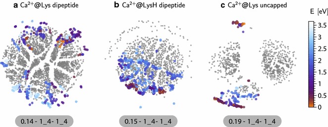
The out-of-sample embedding of conformers with Ca2+ ion on the sketchmap of their pure counterpart, for the three systems we discussed in above: lysine dipeptide (a), protonated lysine dipeptide (b) and molecular lysine (c) systems. The projected conformers are colored with their energy where as the sketchmap on which they are projected are kept all in grey color. The location of the projected conformers allows us to understand how the conformational space of the pure conformers are affected due to presence of the Ca2+ ion