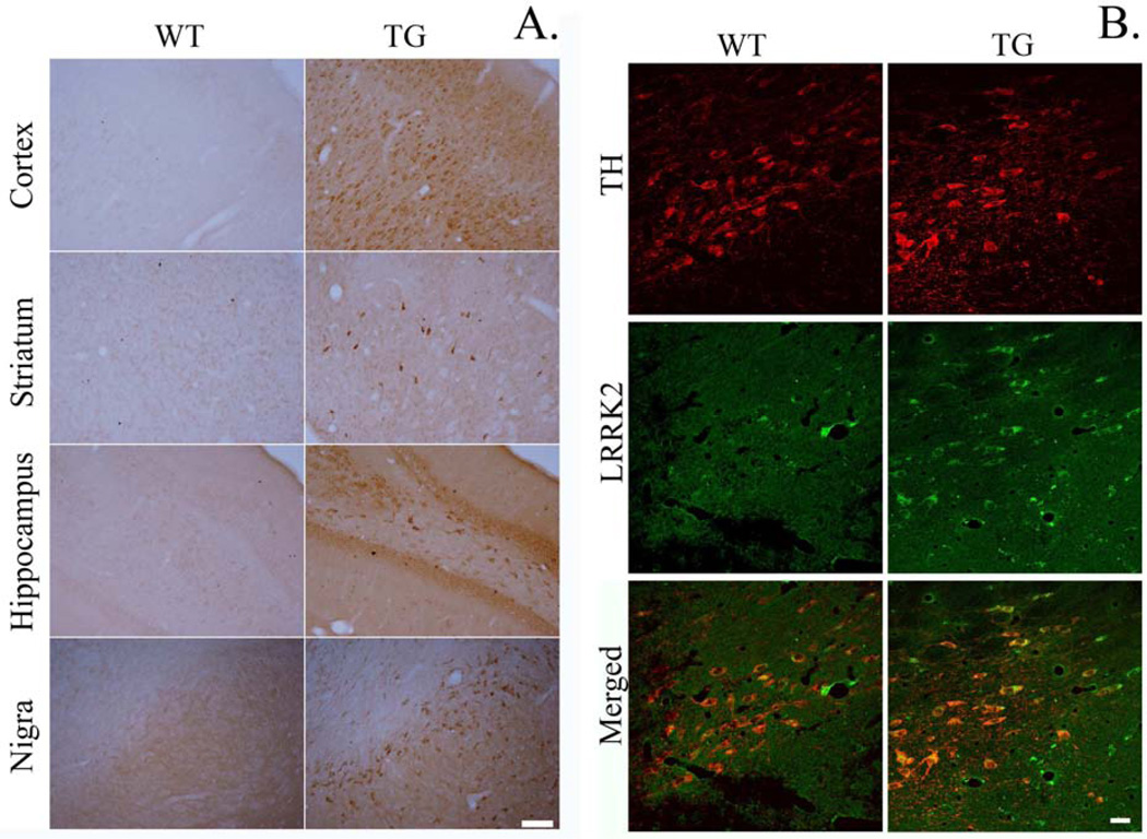
Figure 2.
A. Immunohistochemical staining for LRRK2 shows increased cellular expression in G2019S LRRK2 (TG) compared to wild-type (WT). Coronal brain slices containing cortex, striatum, hippocampus, and substantia nigra were stained for LRRK2. Scale bar = 100 µm. B. Immunofluorescence staining indicated that LRRK2 was highly localized in dopamine (DA) neurons in the SN. Scale bar = 25 µm.

