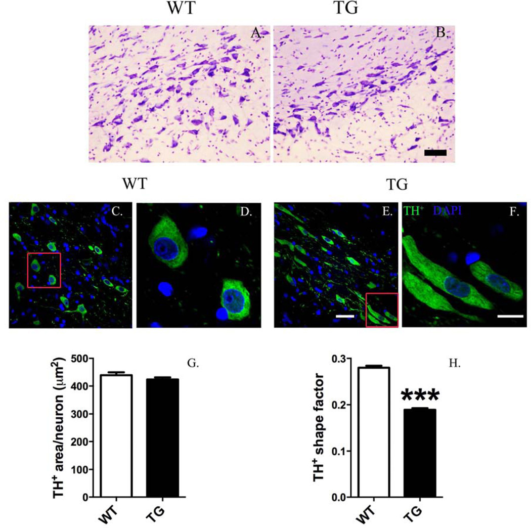
Figure 7.
Dopaminergic (DA) neurons in the substantia nigra of G2019S LRRK2 (TG) rats exhibit an abnormal, elongated morphology. Cresyl violet (Nissl) staining did not reveal gross changes in cellular morphology (scale bar = 50 µm) (A, B). However, quantitative, morphometric analysis of DA neurons revealed changes in cell shape. Low magnification images (A, C) show morphology differences in DA neurons (tyrosine hydroxylase+, TH+) between TG and wild-type (WT) (scale bar = 30 µm). High magnification images (B, D) show that DA neurons in TG rats exhibit an elongated cell body. Morphometric quantification shows that overall cell body area is unchanged (E). Shape area significantly decreased in DA neurons of TG rats (a value from 0 to 1 representing the circularity of an object: a value near 0 reveals a flattened object, whereas a value near to 1 indicates a perfect circle). ***p <0.001, Mann-Whitney U-test versus WT [n = 1016–1220 DA neurons analyzed/group (4 animals/group); DA morphology analyzed at12 months old; data expressed as mean ± S.E.M.].

