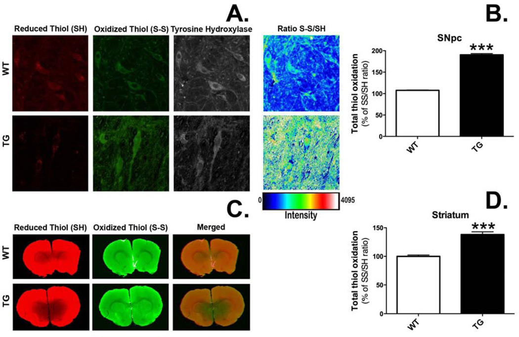
Figure 8.
Rats expressing G2019S LRRK2 (TG) exhibit evidence of increased thiol oxidation in the nigrostriatal dopamine (DA) system. Staining was performed in coronal brain sections for reduced and oxidized thiols in the pars compacta region of the substantia nigra (SN) (A) and the striatum (B). Regions of interest (ROIs) were drawn around single cells in the SN (n = 332 DA neurons/group) or the dorsolateral portion of the striatum (n = 18 striatal ROIs/group). Total thiol oxidation (SS/SH) was determined for each ROI. ***p <0.001 vs. wild-type (WT), Student’s t-test (data expressed as mean ± S.E.M.).

