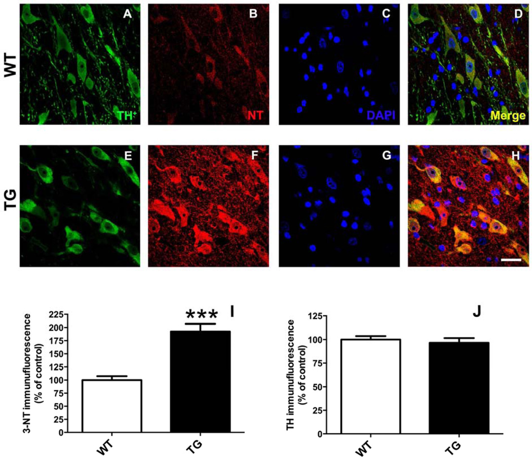
Figure 9.
Rats expressing G2019S LRRK2 (TG) exhibit evidence of increased protein nitrosylation in dopamine (DA) neurons of the substantia nigra (SN). Staining was performed in coronal brain sections for tyrosine hydroxylase (TH) (A, E) and 3-nitrotyrosine (NT) (B, F). Regions of interest (ROIs) were drawn around DAergic neurons (TH+) in the pars compacta region of the SN. Quantitative immunofluorescence is reported (I, J) relative to mean control values (WT). ***p <0.001 vs. WT; n = 5/group; Student’s t-test (data expressed as mean ± S.E.M.). TH levels were not significantly different between groups. Magnification bar = 20 µm.

