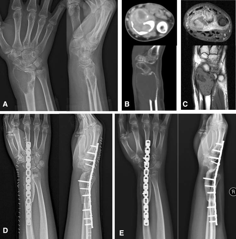
Fig. 1A–E.
A 25-year-old man had GCT in his distal radius on the right side. It was demonstrated in AP and lateral radiographs of the wrist (A), enhanced CT scan (B), and MRI (C) and defined as a Campanacci Grade III GCT. Postoperative AP and lateral radiographic images (D) of the forearm and wrist showed that autogenous structural ICBG was used for reconstruction of the defect created by resection of a distal radius involved by GCT of bone. Six months later, a bony union occurred between ICBG and host bone (metacarpal bone and radius shaft) as shown on AP and lateral radiographs of the forearm and wrist (E).

