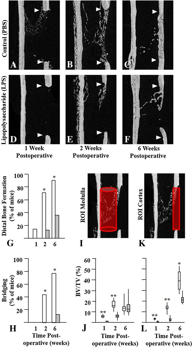
Fig. 1A–L.
Bilateral femoral window defects were generated in skeletally mature mice, and daily intraperitoneal injections of phosphate buffered saline (PBS) or lipopolysaccharide (LPS) were administered for the first 7 days postoperative. Cohorts of mice were euthanized at 1, 2, or 6 weeks postoperative. Femurs were isolated and scanned at a resolution of 5 µm on a Skyscan 1172 microCT instrument. Representative images of the mid-sagittal region with the defects delineated by arrowheads are shown at 1, 2, and 6 weeks postoperative. The upper arrowhead indicates the proximal end of the defect while the lower arrowhead indicates the distal end of the defect. In mice injected with PBS (Control) new bone was seen at the proximal end of the defect at (A) 1 week postoperative, (B) at proximal and distal ends at 2 weeks, and (C) bridging the gap at 6 weeks postoperative. In mice injected with LPS there was little new bone at the (D) proximal end at 1 week or at the (E) distal end at 2 weeks, and (F) most defects failed to bridge by 6 weeks postoperative. (G) Distal bone formation and (H) cortical bridging were decreased in LPS-treated mice (grey bars) compared with Control mice (white bars) at all times. Quantitative analyses of new bone were performed in the (I) medullary canal and in the (K) cortical gap and the results are presented as bar graphs. Bone volume per tissue volume (BV/TV) in the (J) medulla and (L) cortex of LPS-treated mice also was reduced at 1 and 2 weeks postoperative. By 6 weeks postoperative medullary bone was similar but cortical bone remained reduced in the LPS-treated mice compared with Control mice. Results are expressed as median with 25th to 75th quartiles and ranges. Significantly different from LPS-injected mice; *p < 0.01; **p < 0.001; ROI = region of interest.

