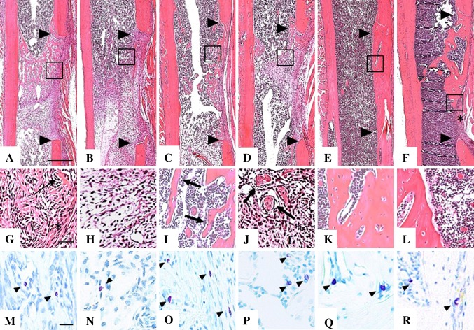
Fig. 2A–R.
Representative images of 5-µm sections from the mid-sagittal region of decalcified, paraffin embedded femurs were stained with hematoxylin and eosin and adjacent sections stained with acidified toluidine blue to identify purple granular mast cells. Low-magnification images of hematoxylin and eosin-stained sections at 1 week for (A) Control and (B) lipopolysaccharide (LPS)-treated mice, 2 weeks for (C) Control and (D) LPS-treated mice, and 6 weeks after surgery for (E) Control and (F) LPS-treated mice show the margins of the defect (arrowheads) and the region of tissue (box) where high-magnification images were taken. At 1 week postoperative (G) Control mice had newly formed bone (arrow) on the endosteal side, and dense, fibrous tissue (asterisk) on the periosteal side of the defect, while (H) LPS-treated mice had only condensed mesenchyme. At 2 weeks postoperative (I) Control femurs showed a clear pattern of bone trabeculae (arrows) surrounded by marrow, and in the defect of (J) LPS-treated mice, disorganized islands of bone (arrows) in mesenchyme are seen. At 6 weeks postoperative the trabecular bone matures into woven bone with a normal cortical shape in the (K) Control group whereas in the (L) LPS-treated group there are large lacunae in the cortical bone and fibrous tissue remaining in the gap (asterisk in Illustration F). Mast cells (arrowheads) appear more numerous in (M) Control than in (N) LPS-treated bones at 1 week postoperative, whereas equivalent numbers were seen at 2 weeks after surgery in the (O) Control and (P) LPS-treated groups. By 6 weeks postoperative mast cell numbers and location were still similar in (Q) Control and (R) LPS-treated mice. Scale bars = 500 µm (A-F), 50 µm (G-L) and 20 µm (M-R).

