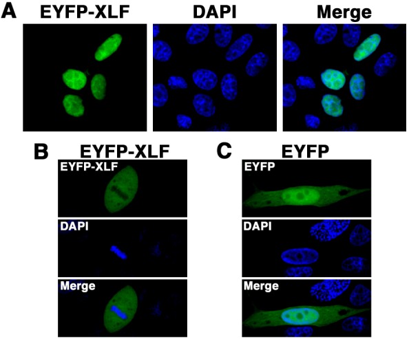
Fig. 3.

Subcellular localization of EYFP-canine XLF in canine cells. Imaging of living EYFP-canine XLF-transfected cells. MDCK cells transiently expressing EYFP-canine XLF (A, B) or EYFP (C) were fixed and stained with DAPI. The stained cells were analyzed by confocal laser microscopy. EYFP images for the same cells are shown alone (left or top panel) or merged (right or bottom panel) with DAPI images (center or middle panel). The images shown are a representative example for interphase cells (C) or mitotic phase cells (B).
