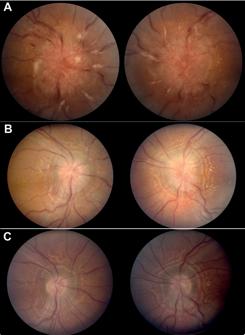
FIGURE 1.
Example of dramatic improvement in optic nerve oedema after optic nerve sheath decompression: (A) pre-operative photographs reveal 4+ optic nerve oedema with associated haemorrhages, cotton wool spots, and exudates; (B) post-operative week 2 photographs reveal resolution of hyperaemia, haemorrhages, and cotton wool spots. In addition, the nerve fibre layer oedema and opacification has diminished; and (C) post-operative week 8 photographs reveal resolution of oedema, hyperaemia, haemorrhages, and cotton wool spots. “High water marks” delineate the region of previous nerve fibre layer oedema. Note: Figure 1 of this article is available in colour online at www.informahealthcare.com/oph.

