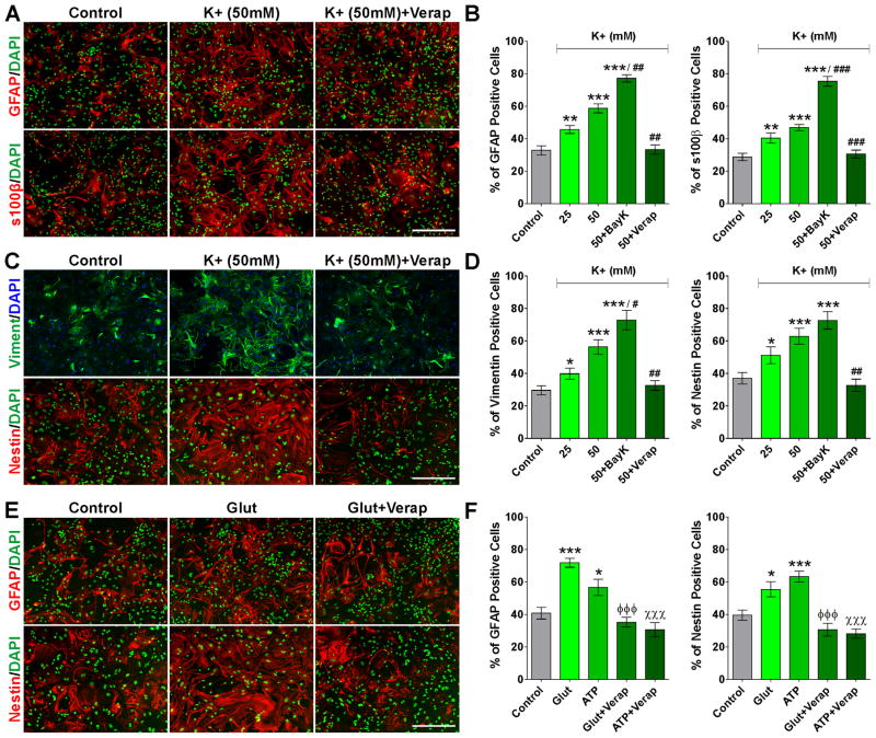
FIGURE 6. High K+, glutamate and ATP induce astrocyte activation in vitro.
After 6DIV, astrocytes were treated with high extracellular K+ (25 and 50mM) for 3 consecutive days and were stained with antibodies against GFAP and s100β (A and B) and vimentin and nestin (C and D). Astrocytes were also treated with glutamate and ATP (10mM) for 3 consecutive days and were stained with antibodies against GFAP and nestin (E and F). Scale bar = 160μm. The percentage of positive cells in each experimental condition was examined by confocal microscopy. Verapamil and Bay K 8644 were applied at 5μM. Values are expressed as mean ± SEM of at least six independent experiments. *p<0.05, **p<0.01, ***p<0.001 vs. control; #p<0.05, ##p<0.01, ###p<0.001 vs. K+ (50mM); ϕϕϕp<0.001 vs. glutamate; χ χχp<0.001 vs. ATP.

