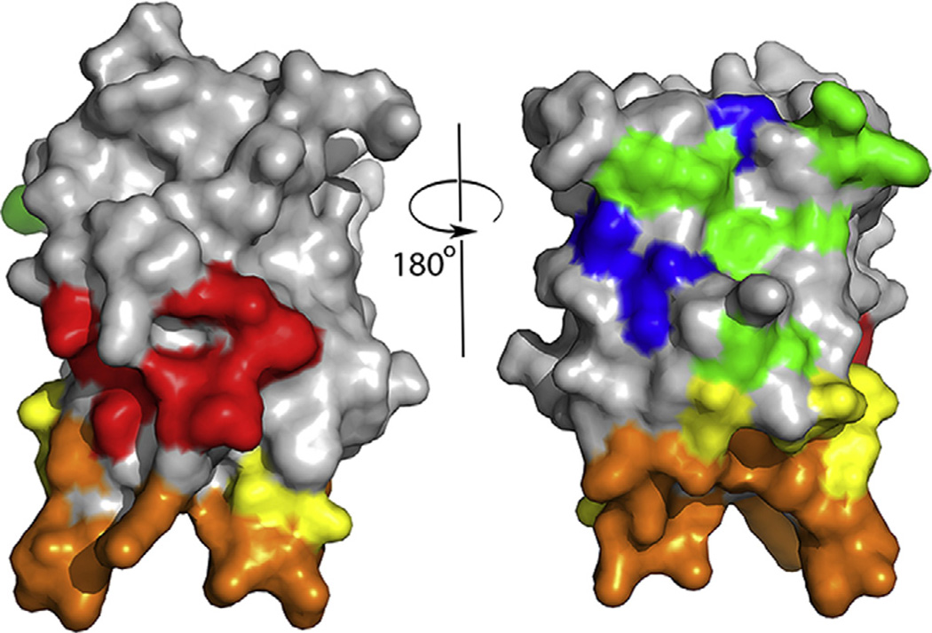
Fig. 1.
Front and back views (rotated 180°) of the FVIII C2 domain crystal structure [37], with surface-exposed amino acids colored to indicate the 5 partially-overlapping B-cell epitopes recognized by 11 neutralizing monoclonal antibodies. The antibodies and epitopes were originally denoted Types A, AB, B, BC and C on the basis of competition ELISA experiments, and the antibodies inhibited distinct binding interactions and functions of FVIII [40]. Thus the identification of specific amino acids comprising these epitopes also indicates which residues and surfaces interact with phospholipid membranes, von Willebrand factor, and components of the intrinsic tenase complex. Type A: red; Type AB: orange; Type B: yellow; Type BC: green; Type C: blue. (For interpretation of the references to colour in this figure legend, the reader is referred to the web version of this article.)

