Figure 3.

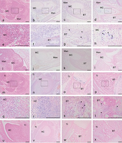
Expression of VEGF-A mRNA based on in situ hybridization with an anti-sense (a-h, m-t) and a sense (i-l, u-x) probe in mouse heads at E 12.5 (a,e,i), E14.5 (b,f,j), E17.5 (c,g,k) and P1 (d,h,l) and tibias at E 12.5 (m,q,u), E14.5 (n,r,v), E17.5 (o,s,w) and P1 (p,t,x). The VEGF-A reaction was localized in (g,h,s,t) (arrowheads); the VEGF-A reaction was scattered in (f,g,r) (arrowheads). e) Magnification of the squared region in (a); f) magnification of the squared region in (b); g) magnification of the squared region in (c); h) magnification of the squared region in (d); q) magnification of the squared region in (m); r) magnification of the squared region in (n); s) magnification of the squared region in (o); t) magnification of the squared region in (p). Mad, mandible; BT, bone trabecula-like matrices; MC, Meckel’s cartilage; TI, tibia; HC, hyaline cartilage. Scale bars: 100 µm. a-d, i-p, u-x) magnification: 10x; e-h, q-t) magnification: 40x.
