. 2017 Jan-Feb;25(1):90–100. doi: 10.1590/1678-77572016-0277

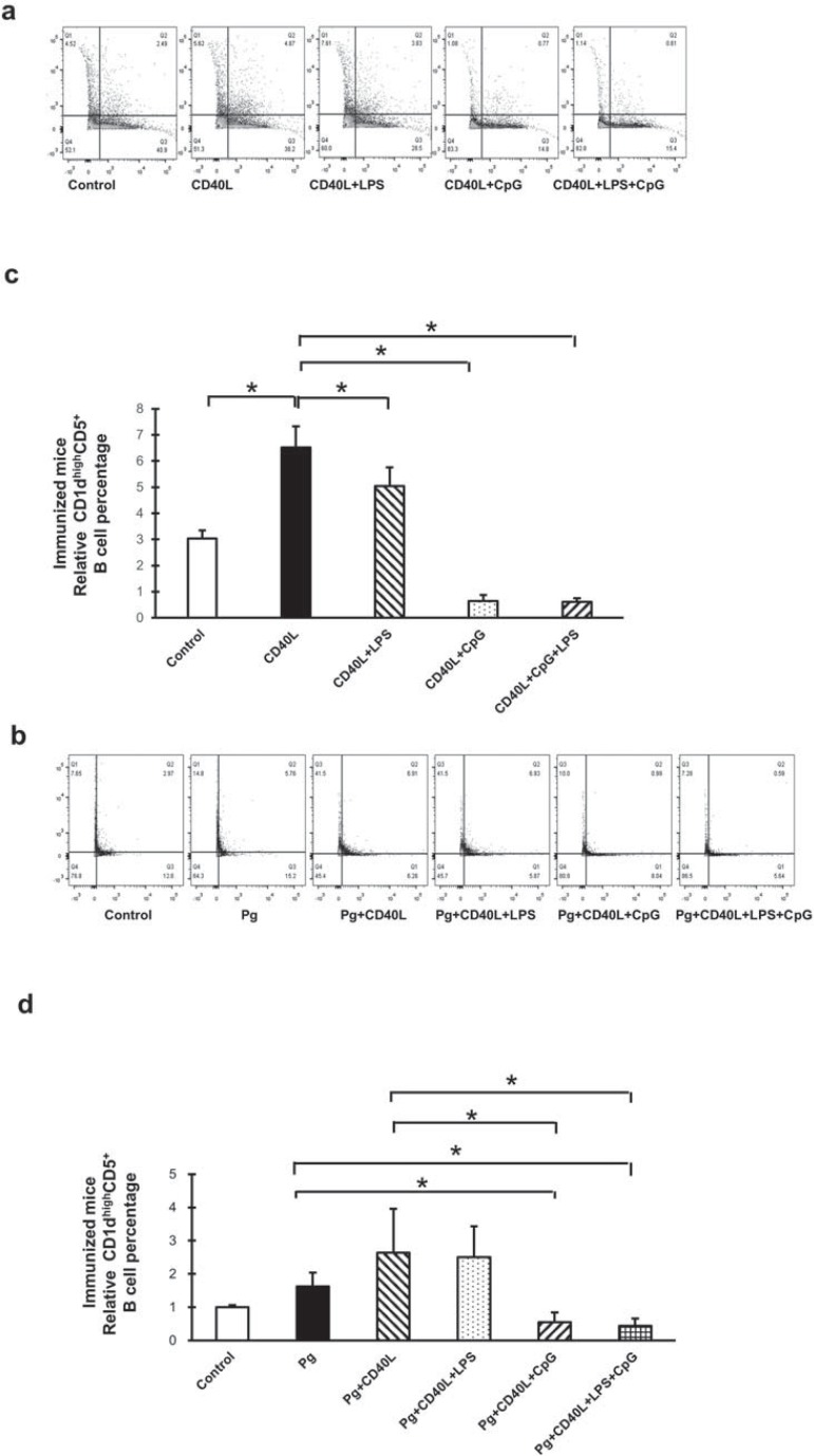
Figure 2. B10 cell expansion in immunized mouse splenocyte B cells after CD40L, LPS, and CpG treatment with/without P. gingivalis co-stimulation. C57BL/6J mice were immunized by intraperitoneal injection of fixed P. gingivalis on day 0 (1×10⁶) and day 7 (1×10⁵). Splenocyte B cells were separated from immunized mice on day 10 and cultured 48 hours with CD40L (1 mg/mL), CD40L (1 mg/mL)+P. gingivalis LPS (10 mg/mL), CD40L(1 mg/mL)+CpG (10 mM), and CD40L (1 mg/mL)+P. gingivalis LPS (10 mg/mL)+CpG (10 mM) in the absence or in the presence of fixed P. gingivalis (5×10⁶per 1×10⁶ cells). CD1highCD5+ B cells were detected using flow cytometry in control and treatment groups without P. gingivalis (a) and with P. gingivalis (b) (X-axis: CD5 PE staining; Y-axis: CD1d APC staining). The percentage of CD1highCD5+ B cells was quantified and analyzed by FlowJo software in control and treatment groups without P. gingivalis (c) and with P. gingivalis (d) (mean±SD, n=3, *p<0.05, detailed statistics information in Table 2).

Figure 2