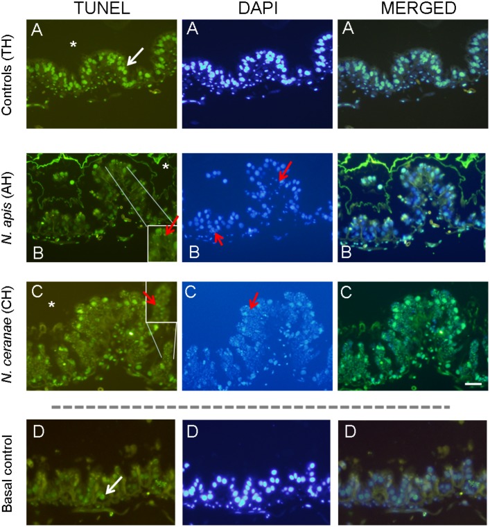
Fig 4. TUNEL assay.
Representative TUNEL / DAPI stained and merged microscopic images of transverse sections from the ventriculum of A: uninfected (controls TH) B: infected with N. apis (AH) or C: N. ceranae (CH) honey bees and treated with cycloheximide. D: Basal apoptosis in uninfected bees and no treated with cycloheximide (Basal control) was very low. The ventriculum cells were counterstained with DAPI (blue). Scale bar = 100 μm. The ventricular lumen is indicated by an asterisk and spores inside ventricular cells are indicated with red arrows.

