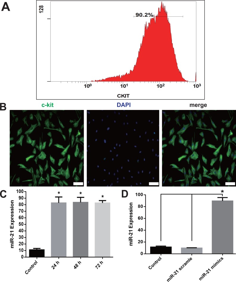
Figure 1. c-kit+ CSCs isolation and overexpression of miR-21in CSCs.
After isolation from rat atrial appendage, cells were purified by a combined use of c-kit antibody and magnetic beads conjugated with secondary antibody. Flow cytometry showed c-kit+ cells were more than 90% (A). (B) Purified cells were double stained by c-kit (green) and DAPI (blue), and observed under a fluorescence microscope (Olympus). Bar = 50 µm. (C) Cultured CSCs were treated with miR-21 mimics for 24, 48 or 72 h before miR-21 RT-PCR detection. miR-21 mimics significantly increased miR-21 but no difference was detected among the three time points. *, P < 0.05 compared with Control. (D) CSCs were incubated with miR-21 mimics or its negative control scramble for 48 h. miR-21 mimics significantly increased miR-21 level in c-kit+ CSCs. *, P < 0.05. n = 3 in each group.

