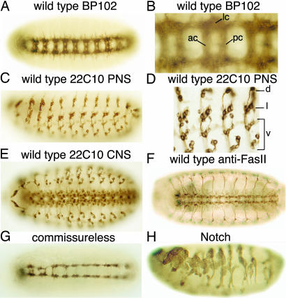
Fig. 1.
Wild-type patterns of the VNC or PNS of Drosophila embryos stained with mAb BP102 (A and B), 22C10 (C-E), or 1D4 (anti-FasII) (F). B and D show high magnifications of A and C, respectively. G shows the VNC of an embryo injected with commissureless dsRNA and stained with BP102. Most commissures are absent. H shows an embryo injected with Notch dsRNA stained with 22C10. Overproduction of PNS neurons and disorganization of the PNS can be seen. Ventral views of embryos are shown in A, B, and E--G; whereas, lateral views are shown in C, D, and H. lc, longitudinal connectives; ac, anterior commissures; pc, posterior commissures; d, dorsal cluster; l, lateral cluster; v, ventral cluster. In all images, anterior is to the left; in lateral views of embryos, dorsal is up.

