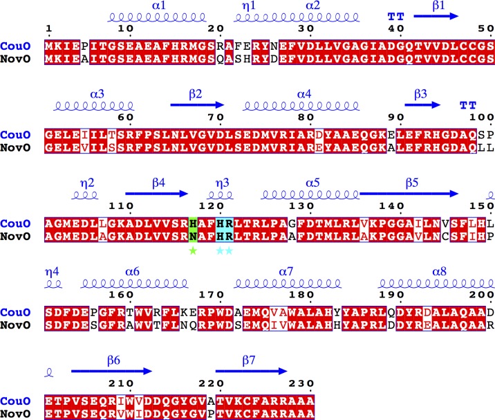
Fig 5. Sequence comparison of NovO and CouO.
Identical residues are shown in red boxes. Secondary structure elements of CouO are shown in blue above the sequence alignment. Important amino acid residues in the active site are marked with an asterisk and shown in cyan (identical) and green (similar) boxes. This figure was prepared using ESPript 3.0 [20].

