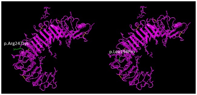
Fig 5. TLR6 exonic SNV positions.
Positions of the exonic SNVs rs5743809 and rs3522046 on the TLR6 protein diagrammed using NCBI Molecular Modeling database (MMDB). The replaced amino acids (p.Leu194Pro and p.Arg247Lys) are shown in green. Variants are located in the extra-cellular domain in the predicted ligand-binding region and may alter hydrogen binding.

