Fig. 5.
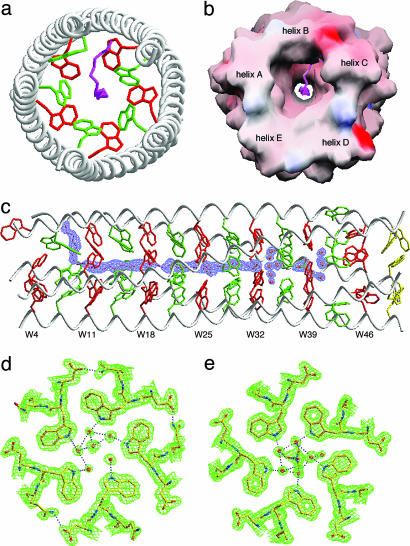
Hydrophobic channel in Trp-14. (a) Axial view of the pentamer. The view is from the N terminus looking down the superhelical axis. The side chains of W7 (d) and W11 (a) are shown in green and red, respectively. A single PEG 400 molecule (pink) is located in the axial channel. (b) Molecular surface representation of the PEG 400 binding site. The solvent-accessible surface is colored according to the local electrostatic potential, ranging from +24 V in dark blue (most positive) to -32 V in deep red (most negative). (c) Side view showing the position of the buried PEG 400 molecule. A simulated annealing omit map covering the bound PEG 400 in a 2Fo - Fc difference Fourier synthesis was calculated with the ligand removed from the model and contoured at 1.0σ. (d) The 2Fo - Fc electron-density map at 1.0σ contour showing the hydrogen bonding network of structured waters in the W32 (a) layer. Water molecules are shown as red spheres, and hydrogen bonds are denoted by pink dotted lines. (e) The 2Fo - Fc electron-density map at 1.0σ contour showing a sulfate ion-mediated hydrogen bonding network of structured waters in the W39 (a) layer. The indole NH group in ≈35% of the Trp residues is hydrogen-bonded in the structure.

