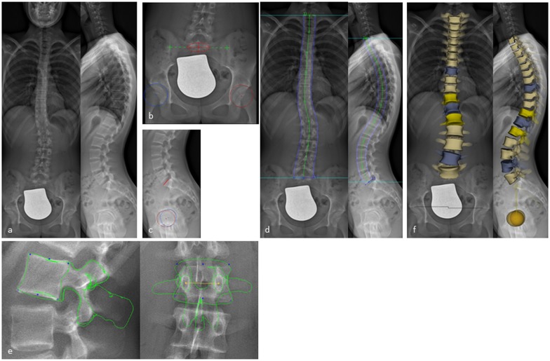
Fig 1. 3D reconstruction from biplanar X-rays using sterEOS.
(a) Biplanar X-rays performed with EOS in a standing position. (b,c) Digitalization of primary anatomical landmarks on the pelvis defining the position of the sacrum (red circle with a cross in the a.p. and red line in the lateral view) and the iliosacral joints (green crosses) as well as on both acetabuli (red and blue circles with a cross in the middle). (d) Shaping the spine from T1 to L5 using green control points. (e) Shaping every vertebral body (T1-L5) by identifying anatomical landmarks using blue and yellow control points on the vertebral bodies resulting in a 3D full spine model (f).

