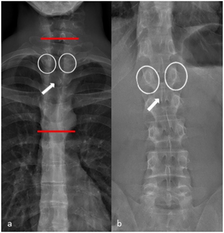
Fig 5. Frontal view X-ray performed with EOS.
(a) Frontal view of the thoracic spine. The pedicles (white circles) and the shape of the processus spinosus (white arrow) are difficult to identify in the upper and middle thoracic spine (area in between the red lines). (b) In the lower thoracic spine and in the lumbar spine identifying the pedicles (white circles) and the shape of the proccessus spinosus (white arrow) is straight forward.

