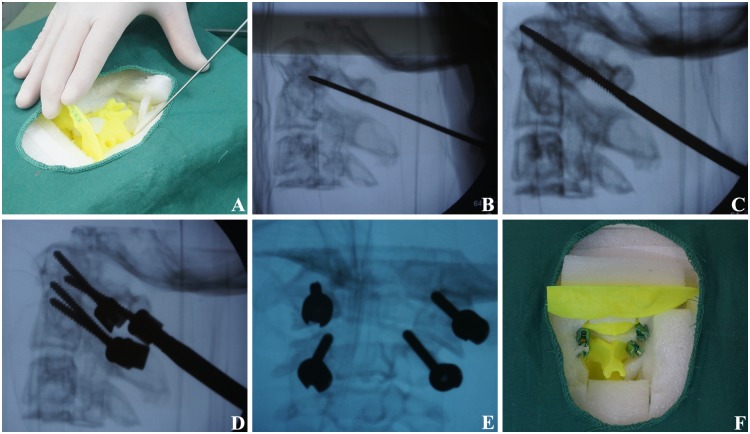
Fig 4. Pedicle screw fixation under C-arm X-ray machine fluoroscopy.
(A) The entrance point was determined according to anatomic marker. (B) The direction of long axis of vertebral pedicle were explored under fluoroscopy. (C) The optimal placement channel was explored under fluoroscopy. (D) The entry angle was adjusted under fluoroscopy. (E) Frontal radiograph was adopted after screw fixation; (F) Pedicle screw fixation was finished.

