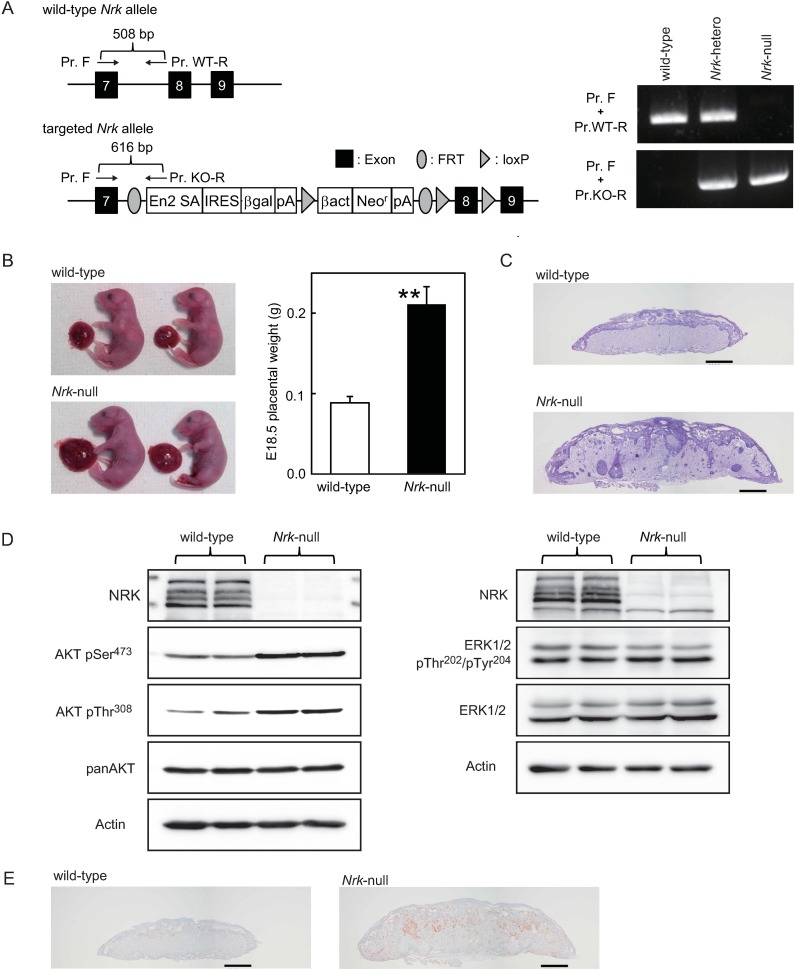
Fig 3. Upregulation of AKT phosphorylation in enlarged placentas derived from Nrk-null E18.5 mouse embryos.
(A) The wild-type Nrk allele and the targeted Nrk allele are shown (left). The arrows indicate PCR primers for detection of each allele. PCR genotyping of mice (right). (B) Wild-type and Nrk-null mouse placentas and foetuses at E18.5 are shown (left). The placental weight was measured at E18.5 in wild-type (n = 24) and Nrk-null (n = 25) embryos (right). Data are presented as mean plus SD. **P<0.01 compared to wild-type. (C) PAS staining of E18.5 placental sections. Scale bars indicate 1 mm. (D) The lysates were prepared from wild-type (n = 2) and Nrk-null (n = 2) placentas at E18.5 and were subjected to western blot analysis. Expressions of NRK, phosphorylated AKT at Ser473, phosphorylated AKT at Thr308 and total AKT were compared between wild-type and Nrk-null placentas (left). Expressions of NRK, phosphorylated ERK1/2 and total ERK1/2 were compared between wild-type and Nrk-null placentas (right). As an internal control, β-Actin was also detected. (E) Immunostaining of E18.5 placental sections with anti-AKT pSer473 antibodies. Scale bars indicate 1 mm. Shown are cropped gels/blots. The gels/blots with indicated cropping lines are shown in S4 Fig.

