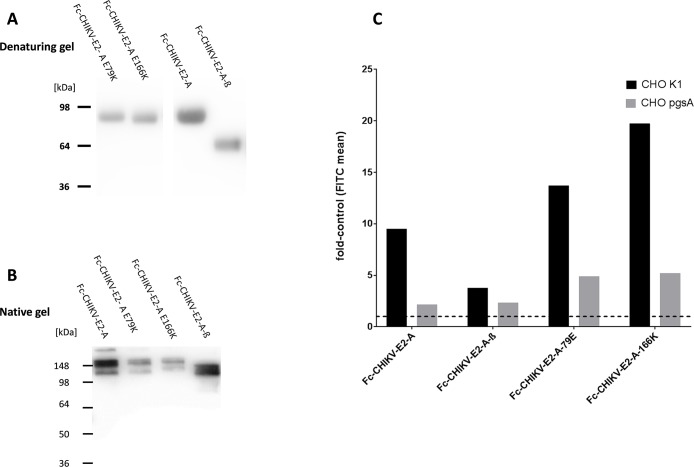
Fig 8. Binding of Fc-fusion proteins containing variants of the CHIKV E2 domain A to CHO-K1 and pgsA-745 cells.
A: Fc-E2 domain A-fusion proteins and Fc protein were expressed from HEK293T cells, affinity purified by protein-A chromatography and separated by SDS-PAGE. The Western blot was detected with an HRP-labeled anti-human IgG antibody. The calculated molecular weights are: Fc-CHIKV-E2-A, E79K and E166K 53 kDa; Fc-CHIKV-E2-A-ß 43,4 kDa. B: Separation of the Fc-fusion proteins under native conditions. The Western blot was detected with an HRP-labeled anti-human IgG antibody. C: CHO-K1 (black) and pgsA-745 (grey) cells were incubated with the indicated recombinant Fc-fusion proteins. Binding was measured by flow cytometry using an anti-human IgG FITC conjugated antibody. The results are shown as fold induction compared to Fc binding. A value of one represents the mean FITC value of the control and is labeled by the dashed line. Values above this line indicate binding. Data represent an average experiment of three independent experiments performed.

