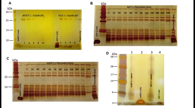
Fig 2. Resistance to trypsin cleavage, pancreatin degradation and pepsin hydrolysis.
Both GLP-1 and mGLP-1 were treated with trypsin (A), pancreatin (Band C) or pepsin (D) at 37°C. The reaction solutions were sampled at different time points and analyzed by SDS-PAGE. Protein bands were visualized using silver staining. mGLP-1 only; mGLP-1 incubated with trypsin for 1, 2, 4, 8, and 12 hours, respectively; GLP-1 only; GLP-1 incubated with trypsin for 1, 2, 4, and 8 hours, respectively. Negative control is trypsin only. GLP-1 (B) and mGLP-1 (C) were incubated with pancreatin for 5, 15, 30, 45, 60, 75, 90, 120, 150, 180, 210, and 240-min, respectively. (D) Lane 1: mGLP-1 incubated with pepsin; Lane 2: mGLP-1 only; Lane 3: GLP-1 incubated with pepsin; Lane 4: GLP-1 only.

