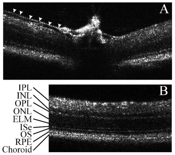
Figure 3.
Large field mouse retina B-scans acquired with a custom built SDOCT. All pictures are single frame pictures without any averaging or smoothing. A) Optic nerve head. Arrow shows a small blood vessel wall, which is different from nerve fiber layer; B) Area near the optic nerve head, showing clear retinal layers including, IPL: inner plexiform layer, INL: inner nuclear layer, OPL: outer plexiform layer, ONL: outer nuclear layer, ELM: external limiting membrane, ISe: external limiting membrane, OS: outer segment, RPE: retinal pigment epithelium.

