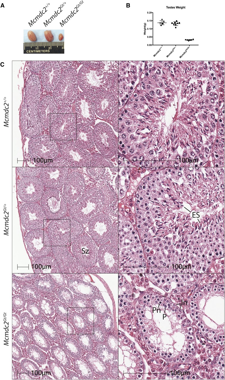
Figure 1.
Testes lacking Mcmdc2 are significantly smaller and lack postmeiotic spermatids. (A) Testes from 12-week-old littermates of the indicated genotypes. (B) Testis weights from Mcmdc2+/+(n = 3), Mcmdc2Gt/+(n = 4), and Mcmdc2Gt/Gt (n = 4) animals. (C) H&E of testes sections from 12-week-old animals of the indicated genotypes. Higher magnification of individual tubules is shown in the insets. The seminiferous tubule enlarged in the lower right panel is representative of the most developmentally advanced observed in mutant testes. As indicated by presence of intermediate spermatogonia and pachytene-like cells (some of which appear necrotic), this appears to be the equivalent of a stage IV tubule (Ahmed and de Rooij 2009). ES, elongated spermatids; In, intermediate spermatogonia; P, pachytene spermatocyte; Pn, likely a necrotic pachytene spermatocyte; Sz, spermatozoa.

