Figure 4.
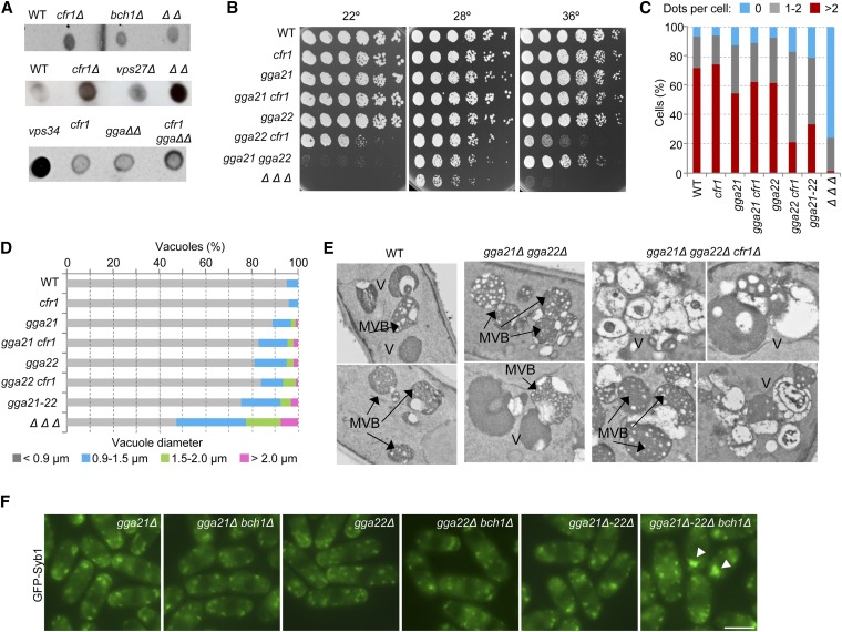
Exomer and GGA adaptors collaborate to maintain the organization of the prevacuolar/vacuolar system. (A) Nitrocellulose membranes, where colonies from the indicated strains had grown at 28°, were analyzed by dot-blot with anti-Cpy1. (B) Cells from the indicated strains were spotted onto YES and incubated at 22° for 7 days, at 28° for 3 days, and at 36° for 2 days. (C) The percentage of cells exhibiting the indicated number of FYVE(EEA1) dots per cells is shown. Dots were scored from photographs in a minimum of 300 cells. (D) Percentage of vacuoles with the indicated diameters. Cells were stained with CDCFDA and photographed. Vacuole diameter was estimated with ImageJ software from the photographs. For each strain, the vacuoles in a minimum of 25 cells were scored. (E) Electron microscopy of the vacuolar system in different strains. Two examples of wild-type and gga21Δ gga22Δ cells and four examples of gga21Δ gga22Δ cfr1Δ cells are shown. (F) GFP-Syb1 distribution. Arrowheads denote fluorescence accumulation inside the cells. Bar, 10 μm. In (C–F), cells were incubated at 32°. In (C), (D), and (F) cells were photographed under a conventional fluorescence microscope. GGA, Golgi-localized, gamma-adaptin ear domain homology, ARF-binding; MVB, multivesicular body; WT, wild-type; YES, 0.5% yeast extract, 3% glucose, 225 mg/l adenine, histidine, leucine, uracil, and lysine hydrochloride, and 2% agar.

