Figure 5.
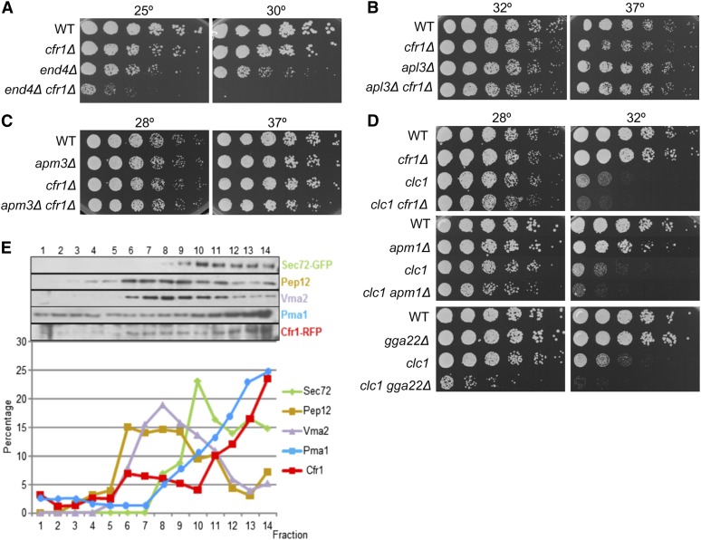
Functional interaction between exomer and other proteins involved in trafficking through endosomes. (A) Cells were spotted onto YES and incubated for 5 days at 25° and for 2 days at 30°. (B–D) The same as in (A), but plates were incubated at the indicated temperatures for 2 days. (E) Cfr1 subcellular distribution. Cell extracts from a strain bearing Cfr1-RFP were centrifuged to equilibrium on mini-step sucrose gradients. Fractions were collected from the lighter to the heavier ones and analyzed by western blot using anti-GFP (Sec72, TGN/EE), anti-Pep12 (PVC), anti-Vma2 (vacuole), anti-RFP and anti-Pma1 (PM) antibodies (upper panel). For each marker, the relative amount in each fraction with respect to its total amount was estimated (lower panel). EE, early endosomes; PM, plasma membrane; PVC, prevacuolar compartment; RFP, red fluorescent protein; TGN, trans-Golgi network; WT, wild-type; YES, 0.5% yeast extract, 3% glucose, 225 mg/l adenine, histidine, leucine, uracil, and lysine hydrochloride, and 2% agar.

